Structure
The pituitary gland, or hypophysis cerebri is a reddish-grey, ovoid body, about 12 mm in transverse and 8 mm in anteroposterior diameter, and with an average adult weight of 500 mg.
Location
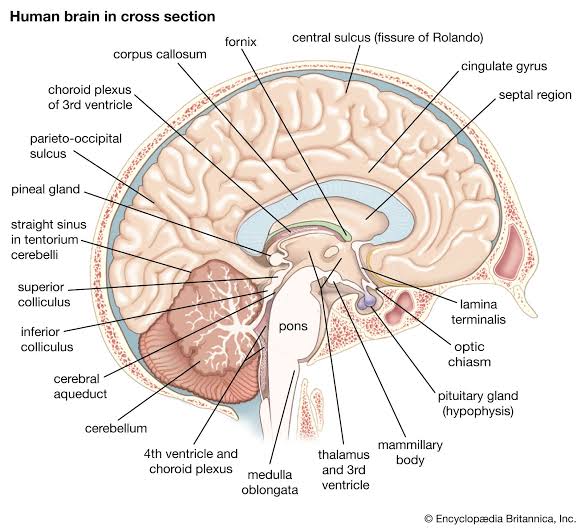
It is continuous with the infundibulum, a hollow, conical, inferior process from the tuber cinereum of the hypothalamus. It lies within the pituitary fossa of the sphenoid bone.

Relations
Superiorly by a circular diaphragma sellae of dura mater. The latter is pierced centrally by an aperture for the infundibulum and separates the anterior superior aspect of the pituitary from the optic chiasma. Inferiorly, the pituitary is separated from the floor of the fossa by a venous sinus that communicates with the circular sinus. The meninges blend with the pituitary capsule and are not separate layers.
Anatomy
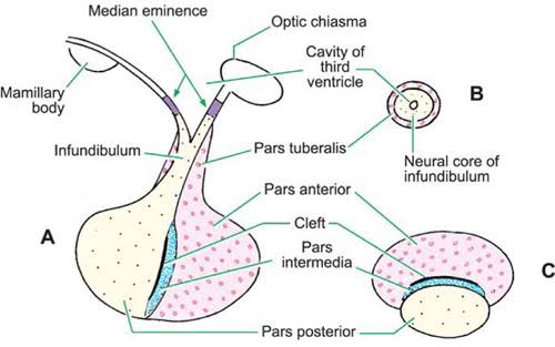
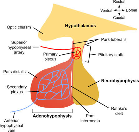
The pituitary has two major parts, the neurohypophysis and adenohypophysis, which differ in their origin, structure and function. The neurohypophysis is a diencephalic down growth connected with the hypothalamus. The adenohypophysis is an ectodermal derivative of the stomatodeum. Both include parts of the infundibulum (whereas the older terms ‘anterior lobe’ and ‘posterior lobe’ do not). The infundibulum has a central infundibular stem, which contains neural hypophysial connections and is continuous with the median eminence of the tuber cinereum. Thus, the term neurohypophysis includes the median eminence, infundibular stem and neural lobe or pars posterior. Surrounding the infundibular stem is the pars tuberalis, a component of the adenohypophysis. The main mass of the adenohypophysis may be divided into the pars anterior (pars distalis) and the pars intermedia, which are separated in fetal and early postnatal life by the hypophysial cleft, a vestige of Rathke’s pouch, from which it develops.

Neurohypophysis
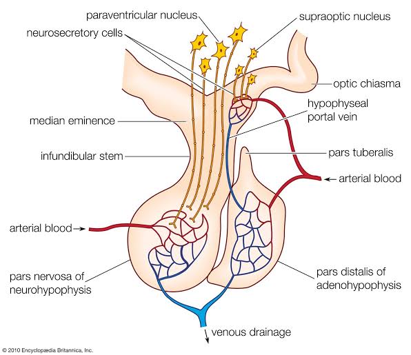
The neurohypophysis includes the pars posterior (pars nervosa, posterior or neural lobe), infundibular stem and median eminence. The adenohypophysis includes the pars anterior (pars distalis or glandularis), pars intermedia and pars tuberalis.In early fetal life, the neurohypophysis contains a cavity continuous with the third ventricle. Axons arising from groups of hypothalamic neurones (e.g. the magnocellular neurones of the supraoptic and paraventricular nuclei) terminate in the neurohypophysis. The long magnocellular axons pass to the main mass of the neurohypophysis. They form the neurosecretory hypothalamohypophysial tract and terminate near the sinusoids of the posterior lobe. Some smaller parvocellular neurones in the periventricular zone have shorter axons, and end in the median eminence and infundibular stem among the superior capillary beds of the venous portal circulation. These small neurones produce releasing and inhibitory hormones, which control the secretory activities of the adenohypophysis via its portal blood supply.
The neurohormones stored in the main part of the neurohypophysis are vasopressin (antidiuretic hormone; ADH), which controls reabsorption of water by renal tubules, and oxytocin, which promotes the contraction of uterine smooth muscle in childbirth and the ejection of milk from the breast during lactation. Storage granules containing active hormone polypeptides bound to a transport glycoprotein, neurophysin, pass down axons from their site of synthesis in the neuronal somata. The thin, unmyelinated axons of the neurohypophysis are ensheathed by typical astrocytes in the infundibulum. Near the posterior lobe, astrocytes are replaced by pituicytes. These are dendritic neuroglial cells of variable appearance, often with long processes running parallel to adjacent axons; they constitute most of the nonexcitable tissue in the neurohypophysis. Typically, their cytoplasmic processes end on the walls of capillaries and sinusoids between nerve terminals. Axons also end in perivascular spaces; they lie close to the walls of sinusoids but remain separated from them by two basal laminae, one around the nerve endings and the other underlying the fenestrated endothelial cells. The spaces between the basal laminae are occupied by fine collagen fibrils.

Adenohypophysis
The adenohypophysis is highly vascular. It consists of epithelial cells of varying size and shape arranged in cords or irregular follicles, between which lie thin-walled vascular sinusoids supported by a delicate reticular connective tissue. Most of the hormones synthesized by the adenohypophysis are trophic. They include the peptides GH, involved in the control of body growth, and prolactin, which stimulates both growth of breast tissue and milk secretion. Glycoprotein trophic hormones are the large pro-opiomelanocortin precursor of ACTH, which controls the secretion of certain suprarenal cortical hormones; TSH; FSH, which stimulates growth and secretion of oestrogens in ovarian follicles and spermatogenesis (acting on testicular Sertoli cells); and LH, which induces progesterone secretion by the corpus luteum and testosterone synthesis by Leydig cells in the testis. Pro-opiomelanocortin is cleaved into a number of different molecules, including ACTH. Beta-lipotropin is released from the pituitary but its lipolytic function in humans is uncertain. Beta-endorphin is another cleavage product released from the pituitary. Neurones that secrete the peptides and amines that control the anterior lobe are widely distributed within the hypothalamus. They are situated mainly in the medial zone, in the arcuate nucleus, medial parvocellular part of the paraventricular nucleus, and periventricular nucleus. Usually obliterated in childhood, remnants may persist in the form of cystic cavities, often present near the adenoneurohypophysial frontier, which sometimes invade the neural lobe. The human pars intermedia is rudimentary. It may be partially displaced into the neural lobe, and has been included in the anterior and posterior parts by different observers. Apart from this equivocation, which is of little significance, the pars anterior and pars posterior may be equated with the anterior and posterior lobes.
When the associated infundibular parts continuous with these lobes are included, the names adenohypophysis and neurohypophysis become appropriate. The epithelial endocrine cells, which secrete the different adenohypophysial hormones, may be distinguished in part by their differing affinities for acidic and basic dyes. Cells staining strongly are described as chromophils, while those with low affinity for dyes are chromophobes. Acidophils stain strongly with acidic dyes, whereas basophils, which are more prevalent in the central part of the gland, stain strongly with basic dyes. Cells may also be classified according to the hormones they synthesize into somatotrophs (GH-secreting acidophils, the most numerous chromophil type); lactotrophs (prolactin-secreting acidophils, which are dominant in pregnancy and hypertrophy during lactation); gonadotrophs (FSH- and LH-secreting basophils); thyrotrophs (TH-secreting basophils); and corticotrophs (ACTH-secreting basophils). Chromophobes are thought to be quiescent or degranulated chromophils, or immature precursor cells, and constitute up to half of the cells of the adenohypophysis. The pars intermedia contains follicles of chromophobe cells that surround cyst-like structures lined by epithelium and are filled, to varying degrees, with glycosylated colloidal material. Secretory products of this region may include cleavage products of pro-opiomelanocortin but their functional significance is uncertain. The pars tuberalis contains a large number of blood vessels, between which are cords or clusters of gonadotrophs and undifferentiated cells. A small collection of adenohypophysial tissue lies in the mucoperiosteum of the human nasopharyngeal roof. By 28 weeks in utero it is well vascularized and capable of secretion, receiving blood from the systemic vessels of the nasopharyngeal roof. At this stage, it is covered posteriorly by fibrous tissue. This is replaced in the second half of fetal life by venous sinuses, and a trans-sphenoidal portal venous system develops, bringing the nasopharyngeal tissue under the same hypothalamic control as the cranial adenohypophysial tissue. The peripheral vascularity of the pharyngeal hypophysis persists until about the fifth year. The organ is then reinvested by fibrous tissue and presumed to be controlled once more by factors present in systemic blood. Though it does not change in size after birth in males, in females it becomes smaller, returning to natal volume during the fifth decade, when once again it may be controlled via a trans-sphenoidal extension of the hypothalamohypophysial portal venous system. The human pharyngeal hypophysis may be a reserve of potential adenohypophysial tissue, which may be stimulated, particularly in females, to synthesize and secrete adenohypophysial hormones in middle age, when intracranial adenohypophysial tissue is beginning to fail.

Arteries and veins of the pituitary gland
The arteries of the pituitary arise from the internal carotid arteries via a single inferior and several superior hypophysial arteries on each side. The former come from the cavernous part of the internal carotid artery, the latter from its supraclinoid part and from the anterior and posterior cerebral arteries. The inferior hypophysial arteries divide into medial and lateral branches, which anastomose across the midline and form an arterial ring around the infundibulum. Fine branches from this circular anastomosis enter the neurohypophysis to supply its capillary bed. The superior hypophysial arteries supply the median eminence, upper infundibulum and, via the artery of the trabecula, the lower infundibulum. (The trabecula is a compact band of connective tissue and blood vessels lying within the pars distalis on either side of the midline; it forms a prominent fibrovascular tuft close to the junction of the central and lateral parts of the pars distalis. A confluent capillary net, extending through the neurohypophysis, is supplied by both sets of hypophysial vessels. Reversal of flow can occur in cerebral capillary beds lying between the two supplies. The arteries of the median eminence and infundibulum end in characteristic sprays of capillaries, which are most complex in the upper infundibulum. In the median eminence, these form an external or ‘mantle’ plexus and an internal or ‘deep’ plexus. The external plexus, fed by the superior hypophysial arteries, is continuous with the infundibular plexus and is drained by long portal vessels, which descend to the pars anterior. The internal plexus lies within and is supplied by the external plexus. It is continuous posteriorly with the infundibular capillary bed and, like the external plexus, is drained by long portal vessels. Short portal vessels run from the lower infundibulum to the pars anterior. Both types of portal vessel open into vascular sinusoids, which lie between the secretory cords in the adenohypophysis and provide most of its blood. There is no direct arterial supply. The portal system carries hormone-releasing factors, probably elaborated in parvocellular groups of hypothalamic neurones, and these control the secretory cycles of cells in the pars anterior. The pars intermedia appears to be avascular.
There are three possible routes for venous drainage of the neurohypophysis: to the adenohypophysis, via long and short portal vessels; into the dural venous sinuses, via the large inferior hypophysial veins; and to the hypothalamus, via capillaries passing to the median eminence. The venous drainage carries hypophysial hormones from the gland to their targets and also facilitates feedback control of secretion. However, venous drainage of the adenohypophysis appears restricted: few vessels connect it directly to the systemic veins and so the routes by which blood leaves remain obscure.
| Hormone | Cell type | Function / effector site | Releasing factor | Inhibitor |
| ACTH | Corticotrophs (basophils) | Suprarenal cortex | CRH | |
| TSH | Thyrotrophs (basophils) | Thyroid gland | TRH | Somatostatin |
| LH, FSH | Gonadotrophs (basophils) | Gonads | GnRH (pulsatile) | GnIH |
| GH | Somatotrophs (acidophils) | Body growth and metabolism | GHRH | Somatostatin |
| Prolactin | Lactotrophs (acidophils) | Mammary gland | Dopamine | |
| Vasopressin (ADH) | Magnocellular hypothalamic neurons | Water absorption in distal renal tubule | ||
| Oxytocin | Magnocellular hypothalamic neurons | Contraction of uterine smooth muscle and ejection of breast milk |


