The oral cavity includes the lips, teeth, tongue, palate, and salivary glands.
Lips
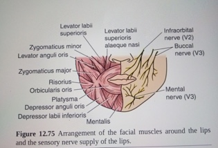
The lips are two fleshy folds that surround the oral orifice. They are covered on the outside by skin and are lined on the inside by mucous membrane. The orbicularis oris muscle and the muscles that radiate from the lips into the face make up the substance of the lips. Also included are the labial blood vessels and nerves, connective tissue, and many small salivary glands. The philtrum is the shallow vertical groove seen in the midline on the superficial surface of the upper lip. Median folds of mucous membrane—the labial frenulae—connect the inner surface of the lips to the gums.

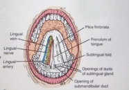
A.Undersurface of the tongue from in front.
B.Oral cavity from in front. The cheek on the left side of the face has been cut away to show the buccinator muscle and the parotid duct.

Arrangement of the facial muscles around the lips and the sensory nerve supply of the lips.
The oral cavity extends from the lips to the pharynx. The paired palatoglossal folds form the oropharyngeal isthmus, which is the entrance into the pharynx. The oral cavity has two components: the vestibule and the oral cavity proper.
Vestibule
The vestibule is a slitlike space that lies between the lips and the cheeks externally and the gums and the teeth internally. It communicates with the exterior through the oral fissure between the lips. When the jaws are closed, it communicates with the oral cavity proper behind the third molar tooth on each side. The vestibule is limited above and below by the reflection of the mucous membrane from the lips and cheeks onto the gums.
The lateral wall of the vestibule is formed by the cheek, which is made up by the buccinator muscle and is lined with mucous membrane. The mucous membrane is tethered to the buccinator muscle by elastic fibers in the submucosa that prevent redundant folds of mucous membrane from being bitten between the teeth when the jaws are closed. The mucous membrane of the gingiva, or gum, is strongly attached to the alveolar periosteum. The tone of the buccinator muscle and that of the muscles of the lips keeps the walls of the vestibule in contact with one another. The duct of the parotid salivary gland opens on a small papilla into the vestibule opposite the upper second molar tooth.
Oral Cavity Proper
The oral cavity proper has a roof and a floor. The palate forms the roof; it consists of the hard palate in front and the soft palate behind. The anterior two thirds of the tongue and the reflection of the mucous membrane from the sides of the tongue to the gum of the mandible largely form the floor. A midline fold of mucous membrane, the frenulum of the tongue, connects the undersurface of the tongue to the floor of the mouth. Lateral to the frenulum, the mucous membrane forms a fringed fold, the plica fimbriata.
The submandibular duct of the submandibular gland opens onto the floor of the mouth on the summit of a small papilla on either side of the frenulum of the tongue. The sublingual gland projects up into the mouth, producing a low fold of mucous membrane, the sublingual fold. Numerous ducts of the gland open on the summit of the fold.
Oral Cavity Sensory Innervation
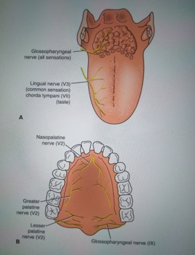
Roof: The greater palatine and nasopalatine nerves from the maxillary division of the trigeminal nerve.

A. Sensory nerve supply to the mucous membrane of the tongue. B. Sensory nerve supply to the mucous membrane of the hard and soft palate; taste fibers run with branches of the maxillary nerve (V2) and join the greater petrosal branch of the facial nerve.
Floor: The lingual nerve (general sensation), a branch of the mandibular division of the trigeminal nerve. The taste fibers travel in the chorda tympani nerve, a branch of the facial nerve.
Cheek: The buccal nerve, a branch of the mandibular division of the trigeminal nerve. Note: the buccal branch of the facial nerve innervates the buccinator muscle, whereas the buccal nerve supplies sensory fibers to the cheek.
Blood supply
The oral cavity and its components receive the blood supply from the facial, the lingual, and the maxillary branches of the external carotid artery. The venous drainage of the oral cavity accompanies its arterial supply, finally draining into the external and internal jugular veins.
The cheeks are supplied by buccaneers branches of maxillary artery.

Lymphatic Drainage
The lymphatic flow from the buccal mucous membrane, the anterior floor of the mouth, the oral tongue, and the hard palate mostly drains toward the submandibular and craniojugular areas.


